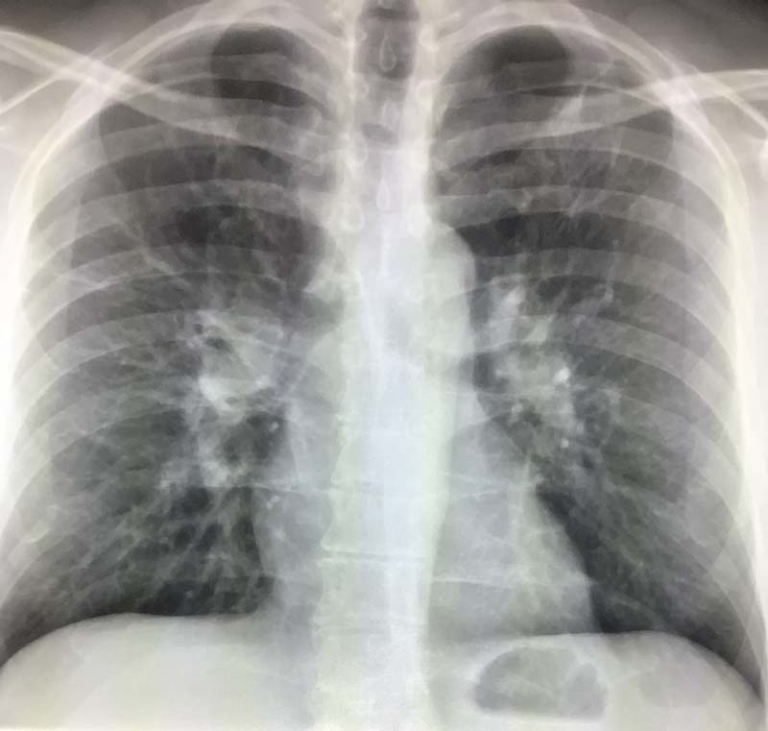
Gastroesophageal Junction
The gastroesophageal junction (GEJ) is the area where the esophagus meets the stomach. It plays a role in digestion and prevents stomach acid from flowing back into the esophagus. When this area is assessed using medical imaging, radiologists look for abnormalities that could indicate conditions such as acid reflux, hiatal hernia, inflammation, or cancer. If you have seen a mention of the gastroesophageal junction on a radiology report, understanding its significance can help you discuss results with your doctor.
How the Gastroesophageal Junction Appears on Imaging
Radiologists evaluate the GEJ using various imaging techniques:
1. X-ray with Barium Swallow
A barium swallow study, also called an esophagram, is a special type of X-ray test that highlights the esophagus and stomach. During this test, the patient drinks a liquid containing barium, which coats the inside of the esophagus and stomach. This contrast material allows radiologists to evaluate the GEJ.
What radiologists look for:
Smooth transition between the esophagus and stomach
Reflux of barium into the esophagus, indicating acid reflux or GERD
Narrowing or irregularity, which could suggest inflammation, scarring, or a tumor
Hiatal hernia, when part of the stomach pushes through the diaphragm into the chest
2. CT Scan of the Abdomen and Chest
A CT scan provides detailed cross-sectional images of the GEJ and surrounding structures. This is often used when doctors suspect complications from reflux, cancer, or infections.
What radiologists look for:
Thickening of the GEJ, which may indicate esophagitis (inflammation) or early cancer
Masses or irregular tissue, which could suggest tumors
Lymph node enlargement, often a concern in cases of esophageal or stomach cancer
Hiatal hernia, seen when part of the stomach moves up into the chest cavity
3. MRI for Soft Tissue Evaluation
MRI is not commonly used for the gastroesophageal junction, but in specific cases, it may provide additional detail, particularly for tumor assessment. Unlike CT, which uses X-rays, MRI uses strong magnets to create detailed soft tissue images.
What radiologists look for:
Tumor invasion into surrounding tissues
Involvement of blood vessels, which helps guide cancer treatment plans
Metastasis or spread of the tumor to other tissues.
4. Endoscopy with Imaging
While not a radiology test, endoscopy is a direct visualization method where a small camera is inserted through the mouth into the esophagus and stomach. It often complements imaging findings by allowing direct tissue biopsy. Some advanced endoscopic techniques use ultrasound (EUS) to assess deeper layers of the GEJ.
What gastroenterologists look for:
Surface abnormalities, such as ulcers, or abnormal growths
Ultrasound findings showing deeper tumor invasion
Tissue samples, taken for biopsy if needed
Common Conditions Affecting the Gastroesophageal Junction
Gastroesophageal Reflux Disease (GERD) and Imaging
GERD is a common condition where stomach acid frequently flows back into the esophagus. Over time, this can cause esophagitis (inflammation) or lead to more serious conditions.
Imaging Findings:
Barium swallow may show acid reflux reaching the esophagus
CT scan may reveal thickening of the esophageal walls
Endoscopy may show inflamed tissue at the GEJ
Hiatal Hernia and How It Appears on Scans
A hiatal hernia occurs when the stomach bulges through the diaphragm into the chest. It is a frequent cause of acid reflux and can be seen on multiple imaging tests.
Imaging Findings:
Barium swallow shows the stomach moving up into the chest
CT scan confirms herniation
MRI (rarely used) may help assess large or complex hernias
Esophageal and Gastroesophageal Junction Cancer
Cancers affecting the GEJ are becoming more common, particularly in people with long-term acid reflux or Barrett’s esophagus (a condition where the esophagus lining changes due to chronic acid exposure).
Imaging Findings:
Barium swallow may show an irregular or narrowed area
CT can sometimes detect tumor and their spread to lymph nodes
MRI helps assess the depth of tumor invasion
Endoscopic ultrasound (EUS) provides a detailed look at tumor involvement
Barrett’s Esophagus and Precancerous Changes
In Barrett’s esophagus, the normal lining of the esophagus changes, increasing the risk of cancer.
Imaging Findings:
CT scan may show thickening of the lower esophagus
Endoscopy is the gold standard, revealing changes in the lining
Biopsy confirms whether precancerous or cancerous cells are present
Why Imaging of the Gastroesophageal Junction Matters
Understanding how the gastroesophageal junction appears on different scans helps doctors diagnose and monitor conditions early. Some issues, like mild acid reflux, may only require lifestyle changes, while others, such as GEJ cancer, need immediate treatment.
What to Do if Your Imaging Report Mentions the GEJ
If your radiology report references the gastroesophageal junction, here are some steps you can take:
Review the report with your doctor to understand the findings.
Ask if additional tests are needed, such as an endoscopy or biopsy.
Discuss lifestyle changes if you have reflux-related issues.
Consider additional imaging if abnormalities were found.
Conclusion
The gastroesophageal junction is where the esophagus meets the stomach. Imaging tests plays an important role in diagnosing conditions affecting it. From barium swallow X-rays to CT scans and endoscopic ultrasound, these tests provide valuable information about reflux, hernias, inflammation, and cancer. If you see the GEJ mentioned on a radiology report, discuss this with your doctor who can combine the imaging findings with your history and suggest the best path forward.
References
1.https://radiopaedia.org/articles/gastro-oesophageal-junction?lang=us
3. https://insightsimaging.springeropen.com/articles/10.1007/s13244-017-0548-3

