Sternotomy
A sternotomy is a surgical procedure where the breastbone (sternum) is cut open to provide access to the heart and other structures in the chest. This procedure is commonly performed during heart surgeries, such as coronary artery bypass grafting, heart valve replacement, or heart transplantation. When reviewing a radiology report that mentions sternotomy, it indicates that the patient has undergone this chest operation, and imaging is showing the post-surgical appearance.
What Does a Sternotomy Look Like on Imaging?
When a radiologist examines images of a patient who has undergone a sternotomy, several characteristic findings appear on various imaging modalities.
X-Ray Findings After Sternotomy
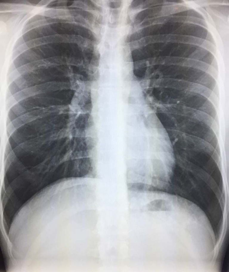
On a chest X-ray, a sternotomy typically appears as a vertical midline density representing the healed or healing sternum. The most distinctive feature is the presence of sternal wires, which appear as bright white horizontal lines crossing the sternum. These wires are used to close the sternum after surgery and remain in the body permanently. The number of wires can vary depending on the surgical technique, but typically ranges from five to eight evenly spaced wires along the length of the sternum.
CT Scan Appearance of Sternotomy
Computed tomography (CT) provides more detailed information about a sternotomy site than X-rays. On CT scans, the sternal wires appear as very bright (high-density) metallic artifacts that can sometimes cause streaking artifacts on the image. CT can reveal important details about the healing process, including:
- The alignment of the sternal edges
- Any gaps between the sternal halves
- Signs of infection or inflammation around the sternotomy site
- Potential complications such as sternal dehiscence (separation of the sternum)
MRI Visualization of Sternotomy
While magnetic resonance imaging (MRI) is less commonly used to evaluate the sternum directly after sternotomy due to artifacts from the metallic wires, it may be performed to assess other chest structures. The sternal wires cause signal void areas (dark spots) on MRI images due to magnetic susceptibility artifacts. Modern MRI techniques can minimize these artifacts to some extent.
Common Radiological Findings Associated with Sternotomy
When examining imaging after a sternotomy, radiologists typically look for several normal and abnormal findings:
Normal Post-Sternotomy Appearance
A normal post-sternotomy appearance includes:
- Well-aligned sternal edges with minimal displacement
- Properly positioned sternal wires
- Gradual healing and fusion of the sternal halves over time
- Minimal soft tissue swelling that decreases over time
In my practice, radiologists typically note normal sternotomy healing as “status post median sternotomy with sternal wires in place and no evidence of complications” on reports when everything appears as expected.
Potential Complications Visible on Imaging
Several complications can be identified on post-sternotomy imaging:
Sternal Dehiscence
Sternal dehiscence refers to the separation of the sternal halves after surgery. On imaging, this appears as a gap between the sternal edges and possibly displaced or broken sternal wires. This is a serious complication that may require surgical intervention. The risk increases in patients with conditions such as obesity, diabetes, or chronic obstructive pulmonary disease.
Sternal Infection
Infection of the sternotomy site, also known as mediastinitis, is a severe complication. Imaging findings may include:
- Fluid collections around the sternum
- Gas bubbles in the soft tissues
- Erosion of the sternal edges
- Widening of the sternal separation
- Surrounding soft tissue inflammation
CT scanning with contrast is particularly valuable for evaluating suspected sternal infections, as it can detect fluid collections that might represent abscesses.
Sternal Fracture or Wire Complications
Sometimes the sternum may fracture around the wires, or the wires themselves may break or migrate. These complications appear on imaging as:
- Discontinuity in the sternum outside the main sternotomy line
- Broken, kinked, or displaced sternal wires
- Unusual positions of wires compared to previous imaging
Long-term Imaging Considerations After Sternotomy
Many patients who have undergone sternotomy will have multiple imaging studies throughout their lives. Long-term radiological considerations include:
Evaluating Sternal Union
Complete healing of the sternum typically takes 3-6 months. Serial imaging can be used to evaluate the progress of sternal union, particularly in patients with delayed healing or symptoms of instability.
Artifact Considerations in Future Imaging
The presence of sternal wires affects future imaging studies:
- Sternal wires may obscure small details in chest X-rays directly behind them
- CT scans may show beam-hardening artifacts around the wires
- MRI studies may have signal voids and distortion near the sternotomy site
Differentiating New Findings from Post-Surgical Changes
One challenge in interpreting imaging after sternotomy is distinguishing normal post-surgical changes from new pathology. Comparison with prior studies is important to identify new or evolving findings that may represent complications or unrelated disease processes.
Conclusion
Understanding sternotomy findings on radiology reports is important for patients who have undergone cardiac surgery. The presence of sternal wires is a permanent reminder of the procedure and will be visible on all future chest imaging. While most sternotomies heal without complications, radiologists play an important role in monitoring the healing process and identifying potential problems such as dehiscence or infection. If your radiology report mentions sternotomy, it simply means the radiologist has noted evidence of this surgical procedure, and in most cases, this represents an expected finding rather than a cause for concern.
References
- https://my.clevelandclinic.org/health/treatments/24016-sternotomy
- https://emedicine.medscape.com/article/1894058-overview
- https://ajronline.org/doi/10.2214/AJR.18.19782