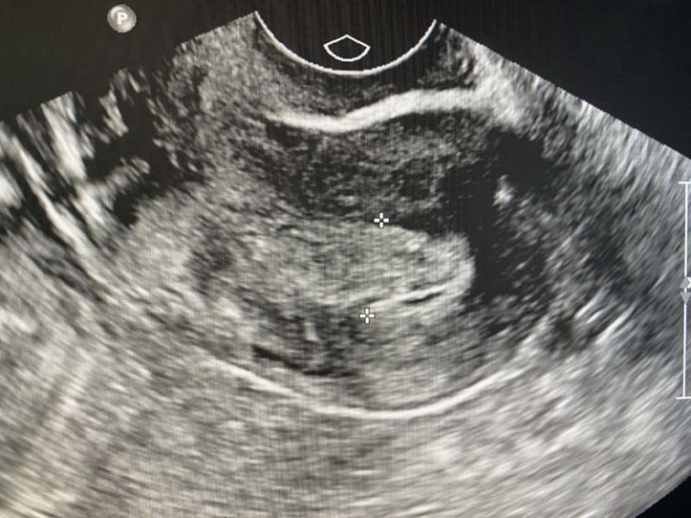
Ectopic Pregnancy Versus Corpus Luteum on Ultrasound
An ectopic pregnancy is a pregnancy that occurs outside the uterus. This is most commonly seen in the Fallopian tubes but can occur in other places like the cervix, ovary and abdomen. Ectopic pregnancy is often diagnosed using ultrasound. Ectopic pregnancy is most commonly seen as a complex cyst or mass next to the ovary in the tube.
A corpus luteum supports the early pregnancy and is seen after ovulation. It’s what’s left in the ovary after the follicle is ruptured and the egg is released into the tube. It usually measures between 1-5 cm and eventually resolves later in pregnancy. It looks like a thick walled complex cyst on ultrasound.
A corpus luteum on ultrasound can mimic the appearance of an ectopic pregnancy. A corpus luteum is within or attached to the ovary which makes an ectopic much less likely. However, sometimes a corpus luteum can be along the periphery of the ovary or abutting it mimicking an ectopic pregnancy.
A technologist can apply transducer pressure to see if the mass moves with the ovary or separate from it. The idea being that the corpus luteum is attached to the ovary and will move with pressure to the ovary. Sometimes it is not possible to exclude an ectopic pregnancy despite our best efforts. Thankfully, close follow up with blood testing and ultrasound can make the diagnosis more confident.
Both an ectopic pregnancy and corpus luteum overlap in the appearance on ultrasound. Both can look like complex cysts with blood flow around them. Ectopics are much more likely to be next to the ovary than within. The location is therefore more important than the actual appearance. Ovarian ectopics are fortunately rare but not excluded.
Both an ectopic pregnancy and corpus luteum can look similar. They can both look like complex cysts. The location is the most important factor. Ectopics occur next to the ovary in the region of the tubes while corpus luteum are most common to be in the ovary. A good ultrasound technologist will be able to distinguish the location of the cyst by applying pressure with the transducer. In some cases, it may not be possible to tell the difference.

