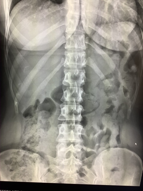
Gastric Antrum
The gastric antrum is the lower most part of the stomach. When you see references to the gastric antrum on a radiology report, it often means there has been imaging done to evaluate this area or that an abnormality has been found. This article will discuss what the gastric antrum is, why it’s significant, and how imaging helps identify abnormalities.
We’ll focus on the role of imaging tests like ultrasound, CT scans, and MRIs in evaluating the gastric antrum. If you’ve ever come across terms like “thickened gastric antrum” or “gastric antral mass” in a report, understanding these findings is important for interpreting results and planning further care.
What Is the Gastric Antrum?
The gastric antrum is the lower part of the stomach. The antrum serves as the last section before the stomach transitions into the small intestine. This area is responsible for breaking down food into smaller particles to prepare it for digestion.
Why Imaging the Gastric Antrum Is Important
Imaging the gastric antrum helps identify several conditions, including ulcers, inflammation, and masses. Radiologists typically assess the gastric antrum during imaging studies for symptoms like abdominal pain, nausea, vomiting, or unexplained weight loss.
Common Reasons for Imaging
1.Chronic Gastritis: Inflammation of the gastric antrum may lead to thickening of its walls, which is can be detectable on imaging.
2.Peptic Ulcers: These can appear in the antrum and are sometimes visible on imaging studies.
3.Tumors: Both benign and malignant masses can develop in the antral region. Imaging may sometimes be used to determine size, location, and extent.
4.Obstructions: A narrowing of the gastric antrum, such as pyloric stenosis, can be identified through specific imaging techniques.
Imaging Techniques for the Gastric Antrum
Radiologists rely on different imaging methods to visualize the gastric antrum. Each modality offers advantages depending on the clinical question and patient presentation.
1. Ultrasound
Ultrasound is a non-invasive, widely available imaging technique that is often used to assess the gastric antrum, especially in pediatric populations. It is particularly useful for detecting pyloric stenosis and monitoring gastric emptying.
•Key Findings on Ultrasound:
•Thickened antral walls may indicate gastritis or inflammation.
•Fluid-filled antral regions could suggest delayed gastric emptying or obstruction.
•Masses or polyps may be seen within the stomach lining.
Personal Insight: In my practice, ultrasound is often used when evaluating suspected pyloric stenosis in infants due to its high sensitivity and lack of radiation exposure.
2. CT Scans
CT scans provide detailed evaluation and are used for evaluating the gastric antrum in adults. They provide cross-sectional images, making it easier to assess surrounding structures and identify complications such as perforations or lymph node involvement.
•Key Findings on CT Scans:
•Thickening of the gastric antrum walls, sometimes with enhancement after contrast, may suggest gastritis or malignancy.
•Submucosal masses, like gastrointestinal stromal tumors (GISTs), can be identified.
•Air-fluid levels in the stomach may point to delayed gastric emptying
3. MRI
MRI is often reserved for complex cases or when additional details about soft tissue are needed. It uses magnetic fields to create detailed images without radiation.
•Key Findings on MRI:
•MRI provides excellent visualization of soft tissue, making it ideal for detecting tumors in the gastric antrum.
•It also evaluates surrounding tissues and blood vessels to determine the extent of any pathology.
What Do Common Imaging Findings Mean?
Thickened Gastric Antrum Wall
This is one of the most frequently reported findings. Wall thickening can be caused by inflammation (gastritis), infection (such as H. pylori), or malignancy.
Gastric Antrum Mass
Masses in the gastric antrum can vary from benign polyps to malignant tumors like adenocarcinoma. Imaging helps determine the nature of the mass, its exact location, and whether it has spread to nearby areas.
Pyloric Stenosis
A narrowed gastric antrum with delayed gastric emptying, often seen in infants, is a hallmark finding for pyloric stenosis. Ultrasound is typically the diagnostic tool of choice for this condition.
Submucosal Lesions
These are growths beneath the lining of the stomach, often detected on CT or MRI. GISTs are a common type of submucosal lesion seen in the gastric antrum.
Preparing for Imaging
To get the most accurate imaging of the stomach:
•Fasting: Most studies require fasting for a few hours beforehand to ensure the stomach is empty.
•Contrast Media: For CT and MRI, a contrast agent may be administered.
•Breath Holding: Patients are often asked to hold their breath during certain imaging sequences to reduce motion artifacts.
When to Consult a Specialist
If your radiology report mentions findings like “thickened gastric antrum” or “gastric antral mass,” it’s essential to consult a gastroenterologist or surgeon. They may recommend additional tests, such as an endoscopy, to directly visualize the gastric antrum and perform biopsies if needed.
Conclusion
Imaging of the gastric antrum with Ultrasound, CT scans, and MRIs can identify abnormalities that help physicians make accurate diagnoses and plan appropriate treatments. While endoscopy is often used for evaluating the stomach, imaging tests can sometimes identify abnormalities non invasively. It is important to consult with your doctor when a report mentions an abnormality in the gastric antrum. They will best know what the next steps for diagnosis and treatment will be.
References:
1.https://pmc.ncbi.nlm.nih.gov/articles/PMC5605014/
2.https://ajronline.org/doi/10.2214/AJR.10.4575
3.https://ctisus.com/learning/exhibit//49005

