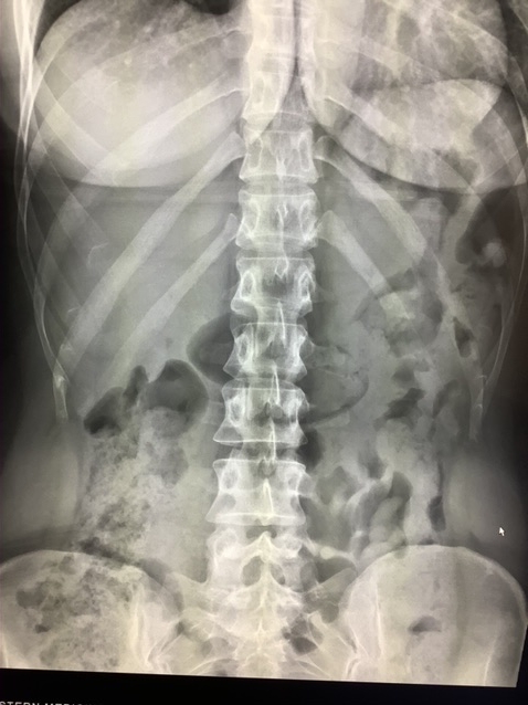
Ileocecal Sphincter
The ileocecal sphincter, also known as the ileocecal valve, plays a role in the digestive system by regulating the flow of material from the small intestine into the large intestine. This small but important structure helps ensure proper digestion and absorption of nutrients while preventing harmful bacteria from entering the small intestine.
Imaging tests are important for diagnosing and understanding conditions related to the ileocecal sphincter. In this article, we’ll explore various imaging tests used to visualize the ileocecal sphincter, along with their advantages and applications.
What is the Ileocecal Sphincter?
The ileocecal sphincter is located between the ileum, which is the last part of the small intestine, and the cecum, the first section of the large intestine. Its primary function is to act as a one-way valve, allowing digested food to pass into the large intestine while preventing backflow. When the ileocecal valve malfunctions, it can result in various gastrointestinal issues, such as bacterial overgrowth or diarrhea.
Imaging Techniques for the Ileocecal Sphincter
1. Abdominal Ultrasound for the Ileocecal Sphincter
One of the first imaging methods used to evaluate the ileocecal sphincter is abdominal ultrasound. This non-invasive procedure uses high-frequency sound waves to produce images of internal organs, including the intestines and sphincters.
Ultrasound is particularly useful for assessing the overall structure of the intestines and detecting any abnormal enlargement or inflammation in the ileocecal area. While the detail provided by ultrasound might not be as high as other imaging methods, it’s often a first-line diagnostic tool due to its simplicity and lack of radiation exposure.
Benefits of Ultrasound:
• Non-invasive and painless
• No radiation exposure
• Cost-effective and widely available
When is Ultrasound Used?
Ultrasound is typically used when there is suspicion of inflammation in the digestive tract or when a non-invasive diagnostic approach is preferred.
2. CT Scans for the Ileocecal Sphincter
A computed tomography (CT) scan is another common method for imaging the ileocecal sphincter. CT scans use X-rays to create detailed cross-sectional images of the body. For the ileocecal sphincter, CT scans can provide high-resolution images, making it easier to detect abnormalities such as thickening of the valve or signs of inflammation in the surrounding tissues. CT scans are particularly valuable when there is a need to evaluate the presence of tumors, blockages, or infections.
Advantages of CT Scans:
• Highly detailed images
• Quick and efficient
• Useful for detecting tumors or blockages
Drawbacks:
• Exposure to radiation
• Not suitable for all patients (e.g., pregnant women)
3. MRI for Ileocecal Sphincter Imaging
Magnetic resonance imaging (MRI) is a more advanced imaging technique that uses powerful magnets and radio waves to create detailed images of the body’s internal structures. MRI provides excellent contrast between different types of tissues, which is particularly useful when imaging the digestive tract and identifying problems with the ileocecal sphincter.
For imaging the ileocecal valve, MRI is effective at showing inflammation, structural abnormalities, and potential obstructions. It is often used when more detail is required than what is available through ultrasound or CT scans.
Why Choose MRI?:
• No radiation exposure
• Highly detailed images of soft tissues
• Can be used to detect inflammation and structural changes
Disadvantages:
• Expensive and less available in some areas
• Longer imaging time, which may be uncomfortable for patients
4. Barium X-rays for the Ileocecal Sphincter
A barium X-ray involves the ingestion or administration of a barium solution that coats the lining of the intestines, making them easier to see on X-rays. While not as commonly used today, barium X-rays can provide important information about the ileocecal sphincter’s function and shape. This technique can help identify problems such as strictures, tumors, or improper function.
Benefits of Barium X-rays:
• Can show movement and function of the intestines
• Helps in identifying structural abnormalities
Limitations:
• Involves radiation exposure
• Barium solution may be uncomfortable for some patients
5. Endoscopic Imaging of the Ileocecal Sphincter
Another useful method for imaging the ileocecal sphincter is endoscopy, where a flexible tube with a camera is inserted through the digestive tract to capture real-time images. In this case, a colonoscopy can be used to reach the ileocecal valve.
Endoscopic imaging is particularly helpful when biopsies are needed to determine if there is an underlying condition affecting the valve, such as Crohn’s disease or cancer. It allows for direct observation and often provides the most accurate diagnosis.
Advantages of Endoscopy:
• Direct visualization of the valve
• Ability to take biopsies for further analysis
Drawbacks:
• Invasive procedure
• Requires sedation in most cases
Common Conditions Diagnosed Through Ileocecal Sphincter Imaging
Imaging the ileocecal sphincter can help diagnose a variety of conditions, including:
• Crohn’s Disease: This inflammatory bowel disease often affects the ileocecal valve, leading to inflammation and strictures. MRI or CT scans are commonly used to detect Crohn’s-related changes.
• Ileocecal Valve Syndrome: A condition where the ileocecal valve becomes dysfunctional, leading to symptoms like bloating, constipation, or diarrhea.
• Ileocecal Tumors: Tumors in the region of the ileocecal valve can be detected through CT scans or MRIs, allowing for early diagnosis and treatment.
• Intestinal Obstructions: CT scans are commonly used to detect blockages at the ileocecal valve.
Conclusion
Imaging is important in the accurate diagnosis and management of conditions affecting the ileocecal sphincter. Techniques like ultrasound, CT scans, MRI, barium X-rays, and endoscopy provide information about the structure and function of the valve. These techniques help to identify issues such as inflammation, tumors, or valve dysfunction.

