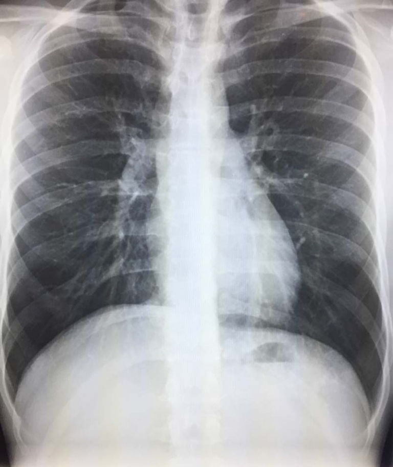
Pneumonia versus cancer on CT scan, can we tell?
It is a common scenario that an abnormal area of the lung on CT can not be clearly diagnosed as a pneumonia or cancer. Sometimes your clinical presentation will favor one over the other, but there is overlap. There is also overlap in the appearance of pneumonia and cancer on imaging studies like CT.
A pneumonia on CT scan will classically be a white area of lung with air filled bronchi traveling through the area. The white lung is the alveoli or air spaces of the lung filled with infectious material. A cancer may look like a round mass, but sometimes it can also either look like a pneumonia or cause a blockage of the air tubes or bronchi which leads to a lung collapse or pneumonia.
The radiologist who interprets your scan will look for findings that favor pneumonia or cancer. Cancer will often block the bronchi or the tubes that carry air to your lung. In this case, the white area of the lung will be solid white without any bronchi going through it. The blood vessels of the lung may also be cut off or blocked by the cancer. Pneumonia doesn’t do that.
Cancer will often be mass like and this can often be seen in the CT scan. If the mass is small, central in the lung, and blocks a bronchus, then all that may be seen is part of the lung collapsing or causing a pneumonia. In some cases, when the CT scan is done without contrast given through your vein, even larger masses may not be seen as they blend in with the other tissues and are hidden.
Cancer which is more peripheral in the lung is often the toughest to tell. Cancer may simply look like a white spot amidst the dark of the normal lung tissues. There may be no secondary clues on imaging or prior studies showing change. The radiologist will look at the edges to see if they are smooth or irregular. Irregular edges are more suspicious. They will look for fat or calcium in the lesion which will suggest a benign diagnosis. But in most cases, you can’t tell simply from the CT scan.
Radiologists will also look for spread of your cancer such as enlarged lymph nodes in your chest. Areas of spread to your bones. Other nodules or spots in the lungs which may mean that the cancer has spread. Often fluid can accumulate in the lung with both pneumonia and cancer. Many of these findings are not seen with pneumonia and will favor cancer.
The radiologist will often make recommendations to try to arrive at the correct diagnosis. A 1-2 month follow up CT may be recommended. The thinking is that pneumonia will clear up after antibiotics and disappear on the scan whereas cancer will not. If the spot in your lung is bigger then 1 centimeter, the radiologist may recommend a PET scan which can help tell if it is suspicious for cancer. If the spot is smaller then 1 cm then a follow up CT may be recommended. In some cases, a biopsy may be recommended which is more definitive.
In some cases you may be referred to a lung specialist (pulmonologist) who will manage your care. In some cases where there is suspicion for cancer, he may do a bronchoscopy which is a test to look directly in the bronchi and sample tissue. Therefore, the imaging is just one part of the workup. There are many options, and there is often no significant delay in the diagnosis which will cause harm.

