Right Subclavian Artery
The right subclavian artery is a major blood vessel that plays an important role in supplying blood to your right arm, shoulder, and parts of your brain. This artery can be mentioned on imaging reports like CT scans. This article will discuss what radiologists look for when examining this artery. This will allow you to work more effectively with your healthcare team.
What Is the Right Subclavian Artery
The right subclavian artery arises from the brachiocephalic trunk which arises from the aorta. This blood vessel runs beneath your right collarbone and serves as the primary pathway for oxygen-rich blood to reach your right arm, shoulder, and hand. The artery also gives rise to several smaller branches that supply blood to muscles in your neck and contribute to brain circulation through the vertebral artery.
Unlike its counterpart on the left side, the right subclavian artery typically originates from the brachiocephalic trunk, also called the innominate artery. This anatomical difference is important for radiologists to recognize when interpreting imaging studies.
Common Imaging Methods for Subclavian Artery Evaluation
When doctors need to examine your right subclavian artery, they have several imaging options available. Each method provides different types of information and has specific advantages depending on what your physician is trying to diagnose or monitor.
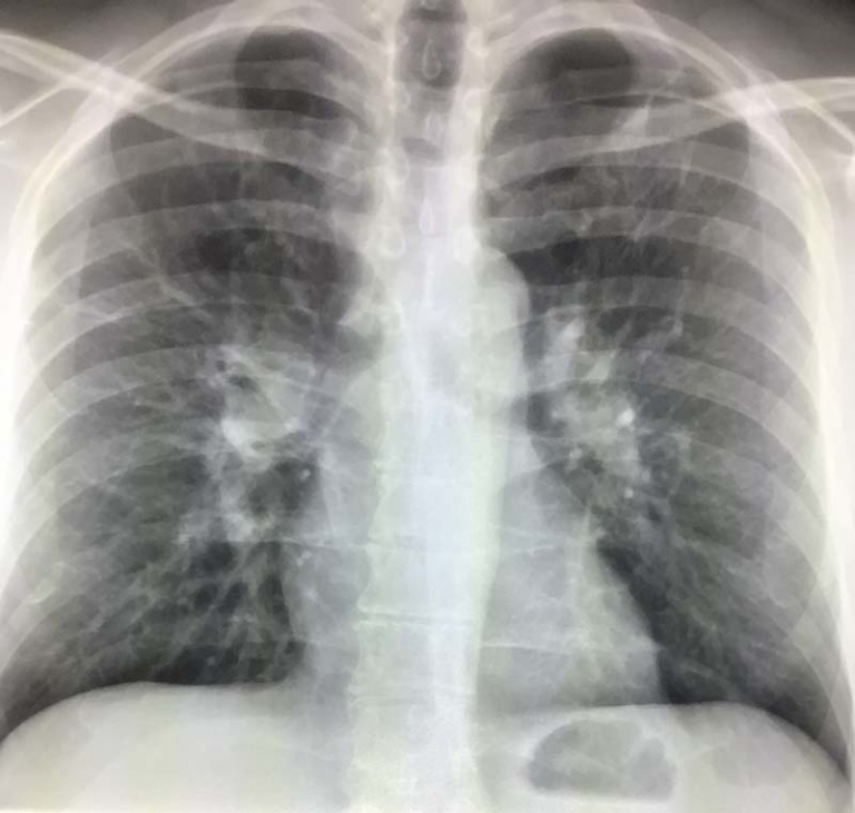
CT Angiography for Subclavian Assessment
Computed tomography angiography, commonly called CT angiography or CTA, is frequently used to visualize the subclavian arteries. This imaging technique involves injecting contrast material into your bloodstream while taking detailed cross-sectional images. The contrast makes your blood vessels appear bright white on the images, allowing radiologists to see the shape, size, and flow patterns within your right subclavian artery.
CT angiography is particularly useful for detecting blockages, narrowing, or unusual branching patterns in the subclavian artery. The test is relatively quick, usually taking less than 30 minutes, and provides excellent detail of both the artery itself and surrounding structures like bones and soft tissues.
MRI and Magnetic Resonance Angiography
Magnetic resonance imaging and its specialized form, magnetic resonance angiography, offer another way to examine the right subclavian artery without using ionizing radiation. MRA can be performed with or without contrast material, making it a good option for patients who cannot receive iodinated contrast used in CT scans.
MRI excels at showing soft tissue details around the subclavian artery and can detect inflammation or other changes in the artery wall that might not be visible with other imaging methods. The technique is particularly valuable for evaluating conditions like thoracic outlet syndrome, where soft tissues may compress the subclavian artery.
Ultrasound Imaging of Subclavian Vessels
Doppler ultrasound provides a non-invasive way to assess blood flow in the right subclavian artery. This imaging method uses sound waves to create real-time images of the artery and can measure the speed and direction of blood flow. Ultrasound is often the first imaging test ordered when doctors suspect problems with the subclavian artery.
The main advantage of ultrasound is that it can be performed quickly in a doctor’s office without any contrast material or radiation exposure. However, ultrasound images of the subclavian artery can sometimes be limited by overlying bone structures or patient body habitus.
Subclavian Artery Abnormalities on Imaging
Radiologists look for several types of abnormalities when examining the right subclavian artery on imaging studies. Understanding these findings can help you better interpret your radiology reports and understand potential treatment implications.
Subclavian Artery Stenosis and Blockages
Narrowing or complete blockage of the right subclavian artery can occur due to atherosclerosis, the same process that affects coronary arteries in heart disease. When radiologists identify subclavian stenosis on imaging, they typically describe the degree of narrowing as mild, moderate, or severe. Severe stenosis may require treatment to restore proper blood flow to the arm. Imaging can also reveal the specific location of narrowing within the subclavian artery, which helps surgeons plan potential interventions.
Subclavian Steal Syndrome on Radiology
Subclavian steal syndrome is a condition where blood flow reverses in the vertebral artery due to significant narrowing or blockage of the subclavian artery. On imaging studies, radiologists can identify this condition by observing reversed flow patterns in the vertebral artery using Doppler ultrasound or specialized MRA techniques.
This condition can cause symptoms like dizziness, arm weakness, or fatigue during arm exercise. Imaging findings of subclavian steal syndrome often prompt further evaluation and potential treatment to restore normal blood flow patterns.
Anatomical Variants and Congenital Differences
The right subclavian artery can have several normal anatomical variants that radiologists encounter on imaging studies. One notable variant is the aberrant right subclavian artery, where the vessel originates directly from the aortic arch rather than from the brachiocephalic trunk.
When radiologists identify anatomical variants, they typically note them in their reports to help other physicians understand the patient’s vascular anatomy. These findings are particularly important if surgical procedures are being planned in the chest or neck area.
Clinical Significance of Subclavian Artery Findings
Understanding what subclavian artery findings mean for your health requires considering both the imaging results and your symptoms. Many people have minor abnormalities in their subclavian arteries that never cause problems, while others may have significant findings that require treatment.
When Subclavian Artery Problems Cause Symptoms
Problems with the right subclavian artery typically cause symptoms in the right arm, including pain, weakness, fatigue, or coolness during physical activity. Some patients also experience numbness or tingling in their fingers. In cases of subclavian steal syndrome, additional symptoms like dizziness or balance problems may occur.
Treatment Options Based on Imaging Results
The treatment approach for right subclavian artery problems depends heavily on imaging findings. Minor narrowing that doesn’t cause symptoms may only require monitoring with repeat imaging studies over time. More significant blockages might need treatment with balloon angioplasty, stent placement, or surgical bypass.
Imaging studies help interventional radiologists and vascular surgeons plan the best treatment approach for each patient. The location, length, and severity of subclavian artery problems all influence treatment decisions, making detailed imaging evaluation essential for optimal care.
Preparing for Subclavian Artery Imaging
If your doctor has ordered imaging studies to evaluate your right subclavian artery, knowing what to expect can help reduce anxiety and ensure the best possible results. Most subclavian artery imaging studies are outpatient procedures that don’t require hospital admission.
For CT angiography, you may need to avoid eating for several hours before the test and should inform your healthcare team about any kidney problems or contrast allergies. MRA typically requires less preparation but may not be suitable for patients with certain metallic implants. Ultrasound studies generally require no special
Conclusion
Understanding imaging results that mention the right subclavian artery is an important step in managing your vascular health. While some findings may seem concerning, many subclavian artery abnormalities can be effectively managed with appropriate medical care.
Working closely with your healthcare team and following their recommendations for treatment and monitoring will help ensure the best possible outcomes for your vascular health. Remember that imaging findings are just one part of your overall health picture, and your doctors will consider these results alongside your symptoms and medical history to develop the most appropriate care plan for your individual situation.
References
- https://my.clevelandclinic.org/health/body/23990-subclavian-artery
- https://www.verywellhealth.com/subclavian-artery-5094725
- https://evtoday.com/articles/2016-nov/aberrant-right-subclavian-arteries