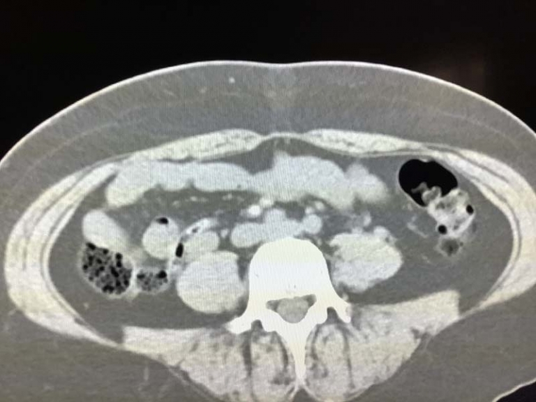
Septic Emboli
Septic emboli indicate an underlying infection that has spread through the bloodstream to another site in the body. These infected clots travel to different organs, most commonly the lungs, brain, and extremities, causing blockages and inflammation. Early detection through imaging is important because septic emboli can lead to severe complications if left untreated.
In radiology, septic emboli appear as small, scattered abnormalities in the affected organ. The most common imaging studies used to detect them include CT scans, MRI, and ultrasound, depending on the location of the suspected emboli.
Septic Emboli in the Lungs: How They Appear on Imaging
The lungs are the most common site for septic emboli, especially in patients with conditions like infective endocarditis or intravenous drug use.
CT Scan Findings
A CT scan is the most effective imaging tool for detecting pulmonary septic emboli. These appear as multiple, small nodules scattered throughout both lungs, often with surrounding areas of inflammation. Some key features include:
- Cavitary Nodules: Some emboli form cavities due to tissue destruction, which is a hallmark of septic emboli.
- Peripheral Distribution: The nodules tend to appear near the outer edges of the lungs rather than the center.
- Feeding Vessel Sign: A small blood vessel can often be seen leading directly to the nodule, indicating the embolic origin.
Chest X-Ray Findings
A chest X-ray is less sensitive but may show vague nodular opacities, which can suggest septic emboli. However, a CT scan is usually needed to confirm the diagnosis.
In my practice, when I see multiple cavitary nodules with a feeding vessel sign on a chest CT, I immediately consider septic emboli, especially in patients with a history of bloodstream infections or intravenous drug use.
Brain Septic Emboli: MRI and CT Findings
Septic emboli can travel to the brain, leading to infections such as brain abscesses or stroke-like symptoms.
MRI Findings
- Multiple Small Infarcts: These are small areas of brain tissue damage due to blocked blood flow.
- Ring-Enhancing Lesions: If the emboli cause an abscess, it may appear as a ring-shaped area on contrast-enhanced MRI.
CT Scan Findings
A non-contrast CT scan may show areas of brain swelling or small hemorrhages. However, an MRI provides more detailed images, making it the preferred method for detecting brain septic emboli.
Septic Emboli in the Extremities: Ultrasound and MRI Findings
Septic emboli can also affect the arms and legs, leading to pain, swelling, and tissue damage.
Ultrasound Findings
- Non-Compressible Veins: If the emboli affect veins, ultrasound may show a clot that does not collapse under pressure.
- Increased Blood Flow on Doppler Imaging: This suggests inflammation and infection.
MRI Findings
- Soft Tissue Swelling and Abscess Formation: MRI with contrast can show infected areas more clearly than ultrasound.
- Bone Involvement: If septic emboli reach the bones, MRI can help detect early osteomyelitis (bone infection).
Common Causes of Septic Emboli
Septic emboli usually originate from infections in the bloodstream. Some common sources include:
- Infective Endocarditis: A heart valve infection that sends clumps of bacteria into circulation.
- Intravenous Drug Use: Bacteria can enter the bloodstream through contaminated needles.
- Septic Thrombophlebitis: A vein infection that releases infected clots.
- Prosthetic Devices and Catheters: Infected medical implants or catheters can introduce bacteria into the bloodstream.
How Radiologists Help Diagnose Septic Emboli
Radiologists play an important role in detecting septic emboli early. When reviewing imaging studies, they look for specific patterns that suggest infection-related emboli rather than other causes like cancer or blood clots. The presence of cavitary nodules in the lungs, ring-enhancing brain lesions, or infected clots in the veins are some of the findings.
Radiologists also compare imaging findings with clinical history. If a patient has a known infection or risk factors like a heart valve issue or IV drug use, radiologists can suggest septic emboli as a likely diagnosis.
Treatment and Prognosis
The treatment of septic emboli depends on the underlying infection. Common approaches include:
- Intravenous Antibiotics: These are essential to clear the infection.
- Surgical Intervention: If there is a heart valve infection or an abscess that does not respond to antibiotics, surgery may be necessary.
- Blood Thinners: In some cases, anticoagulation may be used to prevent further clot formation.
Early detection through imaging significantly improves outcomes. If left untreated, septic emboli can lead to severe complications such as organ failure, stroke, or even death.
Conclusion
Septic emboli indicate a severe bloodstream infection. Imaging plays an important role in identifying these emboli, particularly in the lungs, brain, and extremities. CT scans, MRI, and ultrasound are used to help guide diagnosis and treatment.
References
- https://www.ncbi.nlm.nih.gov/books/NBK549827/
- https://radiopaedia.org/articles/septic-pulmonary-emboli?lang=us
- https://www.medicalnewstoday.com/articles/sepsis-embolism#summary

