TB Chest X-ray
A TB chest X-ray is a vital resource in identifying tuberculosis, a highly contagious lung infection. This article will explore the significance of TB chest X-rays in diagnosing this disease.
Understanding the Purpose:
The purpose of TB chest X-rays lies in their non-invasive and efficient approach to uncovering signs of tuberculosis within the lungs. By capturing chest images, radiologists can detect abnormalities, such as lung nodules, cavities, or infiltrates that might suggest active tuberculosis.
The Procedure:
During a TB chest X-ray, patients stand in front of a specialized X-ray machine while a technician takes a series of images. These images are obtained during breath-holding to ensure the best possible lung visibility.
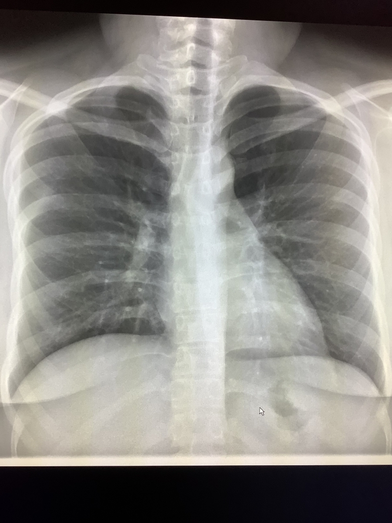
Differentiating Normal and Abnormal Findings:
In normal chest X-rays, clear images with well-defined lung patterns are expected. However, in cases of tuberculosis, you may notice abnormalities. These can include infiltrates, which appear as cloudy areas in the lungs, and cavities, which are hollow spaces.
Distinguishing Active from Latent TB:
TB chest X-rays help distinguish between active and latent tuberculosis. In cases of active TB, X-rays often reveal lung infiltrates, cavities, or enlarged lymph nodes. In latent TB, X-rays may appear normal, though further tests might be necessary to confirm the diagnosis.
The Radiologist’s Report:
After your TB chest X-ray, a radiologist reviews the images and provides a report to your healthcare provider. This report outlines any abnormalities and their locations, enabling precise diagnosis and treatment decisions.
Monitoring and Follow-up:
For patients diagnosed with active tuberculosis, follow-up TB chest X-rays are critical to assess treatment progress. These images aid in understanding if the infection is responding to medication and whether any complications have arisen.
Safety Considerations:
While TB chest X-rays involve a minimal amount of radiation, the benefits of accurate diagnosis and treatment outweigh the risks. Extra precautions are taken for pregnant women and children to minimize radiation exposure.
Complementary Diagnostic Tools:
TB chest X-rays are typically used alongside other tests, such as sputum tests, blood tests, and CT scans, to ensure a comprehensive evaluation of the patient’s condition.
Key Takeaways:
In summary, TB chest X-rays are invaluable in the diagnosis of tuberculosis. They offer a visual representation of the lungs, enabling healthcare professionals to identify signs of active tuberculosis and monitor treatment efficacy. Early detection and treatment are crucial in managing tuberculosis effectively.
Conclusion:
If you suspect you may have tuberculosis or have been in contact with an infected person, seek guidance from a healthcare provider without delay. They can determine if a TB chest X-ray is necessary and guide you through the diagnostic process. In the fight against tuberculosis, early diagnosis is essential, and TB chest X-rays are an indispensable tool.