Mediastinal Lymph Nodes
Radiology reports of the chest can sometimes mention “mediastinal lymph nodes”. These nodes are a normal part of your immune system but can be mentioned when enlarged or abnormal in appearance. Understanding what they are, how they appear on imaging, and what radiologists look for can help you better understand your radiology report.
What Are Mediastinal Lymph Nodes?
Mediastinal lymph nodes are small, oval-shaped structures located in the mediastinum, the central part of the chest between the lungs. They play an important role in filtering harmful substances, fighting infections, and helping the body respond to disease. These lymph nodes are common sites of inflammation and can enlarge due to infections, cancer, or immune disorders.
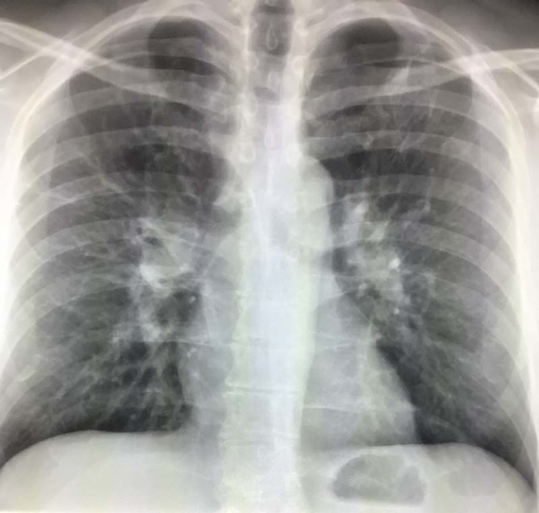
How Mediastinal Lymph Nodes Appear on Imaging
Mediastinal lymph nodes are typically evaluated using:
- Chest X-ray – May show enlarged lymph nodes but lacks the detail to assess their size and appearance accurately.
- CT scan (Computed Tomography) – The most effective imaging test for evaluating mediastinal lymph nodes, showing their size, shape, and any abnormal features.
- PET scan (Positron Emission Tomography) – Used in cases of suspected cancer, as it detects increased metabolic activity in lymph nodes.
- MRI (Magnetic Resonance Imaging) – Occasionally used, when CT is contraindicated or when more detailed analysis is needed.
Normal vs. Enlarged Mediastinal Lymph Nodes
In radiology, a normal mediastinal lymph node is typically less than 10 mm (1 cm) in short-axis diameter on a CT scan. When nodes become larger, radiologists describe them as enlarged or lymphadenopathy, which may require further evaluation.
Causes of Enlarged Mediastinal Lymph Nodes
There are many reasons mediastinal lymph nodes may appear larger than normal on imaging. Some common causes include:
- Infections – Pneumonia, tuberculosis, and viral infections like COVID-19 can cause lymph nodes to swell.
- Inflammatory Conditions – Sarcoidosis, an immune disorder, often leads to enlarged mediastinal nodes.
- Cancer (Malignancy) – Lung cancer, lymphoma, and metastatic disease can cause lymphadenopathy.
- Benign Reactive Enlargement – Sometimes, lymph nodes enlarge temporarily due to minor illnesses without indicating serious disease.
When Are Enlarged Mediastinal Lymph Nodes Concerning?
Radiologists assess multiple factors when evaluating enlarged mediastinal lymph nodes:
- Size – Nodes larger than 1 cm are more suspicious, but not always abnormal.
- Shape – Round nodes may suggest malignancy, while elongated ones are more often reactive.
- Distribution – Widespread lymphadenopathy may indicate cancer or systemic disease.
- Density – Calcified nodes often indicate past infections like tuberculosis.
- Metabolic Activity – PET scans highlight “hot” nodes, which may require biopsy if cancer is suspected.
How Radiologists Report Mediastinal Lymph Nodes
When reading a CT scan, radiologists describe mediastinal lymph nodes based on their:
- Location – Certain regions of the mediastinum (such as paratracheal, subcarinal, or hilar) are more likely to show enlarged nodes in specific diseases.
- Size and Number – Multiple enlarged nodes raise more suspicion than a single slightly enlarged node.
- Appearance Over Time – Comparing current and prior imaging helps determine if changes are new or stable.
Personal Insight: What I Look for in My Practice
In my practice, I often see mildly enlarged mediastinal lymph nodes that turn out to be benign reactive changes. However, when nodes are very large, clustered, or growing over time, further workup with a PET scan or biopsy may be needed. Patients often worry about the term “lymphadenopathy,” but it’s important to remember that not all enlarged nodes mean cancer.
Next Steps After Finding Enlarged Mediastinal Lymph Nodes
If your radiology report mentions mediastinal lymphadenopathy, your doctor may recommend:
- Follow-up Imaging – A repeat CT scan in a few months can track changes.
- Blood Tests – Checking for infections or immune conditions.
- Biopsy – If cancer is suspected, a tissue sample may be taken via bronchoscopy or mediastinoscopy.
Conclusion
Mediastinal lymph nodes are an important part of the body’s immune system, and their abnormal appearance on imaging doesn’t always signal a serious problem. Radiologists evaluate their size, shape, and other features to determine whether further testing is necessary. If your report mentions them, your doctor will guide you on the next steps after considering your medical history and the imaging findings.
References
1. https://www.medicalnewstoday.com/articles/mediastinal-lymphadenopathy#treatment
2. https://www.ncbi.nlm.nih.gov/books/NBK532863/PET-CT in the Assessment of Mediastinal Lymph Nodes
3.https://radiopaedia.org/articles/mediastinal-lymph-node-enlargement?lang=us