Parapneumonic Effusion
Parapneumonic effusion often develops in patients with pneumonia. It refers to the accumulation of fluid in the pleural space due to an infection in the lungs. Radiologists play an important role in detecting and characterizing parapneumonic effusions. This article focuses on how parapneumonic effusion appears on different imaging modalities, key radiologic signs to look for, and what these findings mean for patient management.
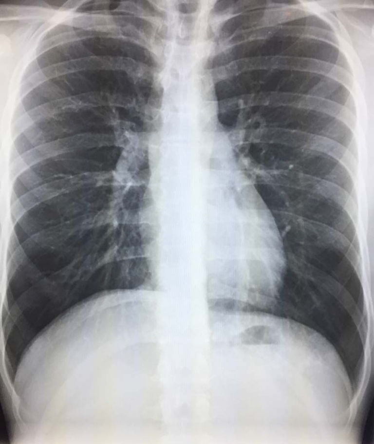
Chest X-Ray Findings in Parapneumonic Effusion
Blunting of the Costophrenic Angle
One of the earliest signs of a parapneumonic effusion on a chest X-ray is blunting of the costophrenic angle. Normally, this angle should be sharp and well-defined. When fluid accumulates, it causes a smooth, concave opacity at the lung base.
Meniscus Sign
A classic radiologic feature of parapneumonic effusion is the meniscus sign. The fluid creates a curvilinear interface with the lung, rising higher along the lateral chest wall than in the center. This helps differentiate it from other causes of lung opacities.
Lateral Decubitus View for Small Effusions
A standard frontal X-ray may miss small parapneumonic effusions. In these cases, a lateral decubitus X-ray, where the patient lies on their side, can confirm the presence of free-flowing fluid. If the fluid shifts, it indicates a simple effusion rather than a loculated collection.
Ultrasound in Parapneumonic Effusion: A Real-Time Tool
Anechoic or Complex Fluid Collection
Thoracic ultrasound is sensitive for detecting parapneumonic effusions, even when X-rays appear normal. Simple effusions appear as anechoic (black) fluid, while complex or infected effusions show internal echoes, septations, or fibrin strands.
Evaluation of Septations and Loculations
Bedside ultrasound often helps determine whether an effusion is simple or complicated. Complicated parapneumonic effusions contain septations, which appear as thin linear echoes within the fluid.
Guidance for Thoracentesis
Radiologists and pulmonologists frequently use ultrasound to guide thoracentesis, a procedure to drain pleural fluid. Identifying the best site for needle insertion improves safety and reduces complications.
CT Scan: The Gold Standard for Parapneumonic Effusion
Fluid Density and Contrast Enhancement
A CT scan provides the most detailed evaluation of parapneumonic effusions. Fluid collections appear as low-density areas in the pleural space. After contrast administration, enhancement and thickening of the pleural lining suggests an inflammatory process.
Split Pleura Sign: A Key Indicator of Infection
One of the most important CT findings in complicated parapneumonic effusion is the split pleura sign. This occurs when the visceral and parietal pleura appear thickened and contrast-enhancing, indicating inflammation. This sign is highly suggestive of empyema, a serious complication that requires drainage.
Loculated Effusions vs. Free-Flowing Fluid
CT also helps differentiate between free-flowing and loculated effusions. While simple parapneumonic effusions tend to distribute evenly, loculated effusions appear as irregular, non-dependent fluid collections, often due to adhesions within the pleural space.
Key Imaging Differences: Parapneumonic Effusion vs. Empyema
Distinguishing between a parapneumonic effusion and empyema is important because treatment approaches differ.
Parapneumonic effusions are usually free flowing, don’t have septations, are have homogeneous fluid
Empyemas are often loculated, have septations, thickened pleura, may have air fluid levels.
When to Be Concerned: Signs of Complication
Rapidly Increasing Effusion Size
A rapidly enlarging pleural effusion raises concern for empyema or hemorrhage. Follow-up imaging is crucial to monitor changes.
Air Bubbles or Gas in the Effusion
If air bubbles or gas pockets are seen within the effusion on CT, this suggests bacterial infection or a bronchopleural fistula, both of which require intervention.
Mediastinal Shift
Large effusions may cause mediastinal shift, where the heart and trachea are pushed away from the affected side. This can impair lung function and require drainage.
Conclusion
Parapneumonic effusion is a frequent complication of pneumonia that radiologists can detect using chest X-rays, ultrasound, and CT scans. Each imaging modality provides additional information from simple effusions to complex infections requiring intervention. Recognizing key radiologic signs such as the meniscus sign and split pleura sign helps with diagnosis and improves patient outcomes.
References
1.https://www.ncbi.nlm.nih.gov/books/NBK534297/
3.https://www.sciencedirect.com/science/article/pii/S0954611121004145

