Abberant Subclavian Artery
An aberrant subclavian artery is a vascular anomaly that occurs during fetal development, affecting the major blood vessels in the chest. This condition may go unnoticed for years, causing no symptoms, but when symptoms arise or for diagnostic purposes, various imaging techniques play an important role in its identification and assessment.
What is an Aberrant Subclavian Artery?
An aberrant subclavian artery, also known as arteria lusoria, is a vascular anomaly where the artery supplying blood to the right arm arises from the aortic arch in an atypical location. Instead of arising as a branch of the brachiocephalic artery, the right subclavian arises as a separate branch directly from the aortic arch. The vessel then courses back to the right side.
Types of Imaging for Diagnosis
Imaging techniques are important in identifying and understanding the complexities of an aberrant subclavian artery. They provide detailed insights into the anatomy, course, and potential complications associated with this vascular anomaly. Let’s discuss these imaging modalities:
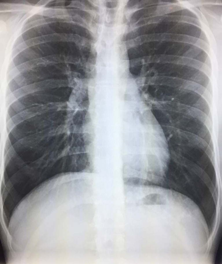
1. X-Ray Imaging:
X-ray imaging utilizes low doses of radiation to produce images of the body’s internal structures. In cases of an aberrant subclavian artery, X-rays might reveal abnormalities or an elongated shadow caused by the altered positioning of the artery. However, X-rays do not offer detailed information on the exact anatomical course or the potential compression of adjacent structures.
2. Computed Tomography (CT) Scan:
CT scans combine X-rays with advanced computer processing to generate detailed cross-sectional images of the body. In diagnosing an aberrant subclavian artery, CT scans are particularly useful. They provide clear and precise visualizations, highlighting the exact origin, course, and relationship of the artery with neighboring structures. CT imaging assists in identifying any compression of the trachea, esophagus, or nerves caused by the aberrant artery.
3. Magnetic Resonance Imaging (MRI):
MRI uses powerful magnets and radio waves to create highly detailed images of soft tissues within the body. In the context of an aberrant subclavian artery, MRI offers a radiation-free alternative to CT scans. It provides excellent anatomical details, helping visualize the course and potential compression of nearby structures caused by the aberrant artery. Additionally, MRI aids in assessing blood flow patterns and identifying associated complications.
4. Angiography:
Angiography involves injecting a contrast dye into the blood vessels and using X-rays to visualize the arterial system. This imaging technique is particularly valuable for mapping the precise course and branches of the aberrant subclavian artery. Angiography provides real-time images of blood flow and helps in detecting any associated anomalies or complications, aiding surgeons in planning interventions accurately.
Importance of Imaging in Diagnosis and Treatment Planning
Accurate imaging of an aberrant subclavian artery is critical for several reasons:
- Precise Diagnosis: Imaging techniques help in confirming the presence of an aberrant subclavian artery, ruling out other conditions, and determining its exact course.
- Assessment of Complications: These imaging modalities can identify any compression of nearby structures, such as the trachea or esophagus, guiding treatment decisions.
- Surgical Planning: For cases requiring surgical intervention, detailed imaging assists surgeons in planning and executing procedures, minimizing risks and complications.
Challenges in Imaging
While various imaging techniques exist, there can be challenges in visualizing an aberrant subclavian artery due to its anatomical variation and the possibility of overlapping structures on some modalities. Specialized imaging protocols and experienced radiologists are crucial to accurately identify and assess this anomaly.
Conclusion
Imaging plays an important role in identifying and evaluating an aberrant subclavian artery. Through X-ray, CT scans, MRI, and angiography, medical professionals can precisely diagnose this vascular anomaly, assess potential complications, and plan appropriate treatments. Early detection and accurate imaging are essential for optimal management, ensuring better outcomes for individuals affected by this condition.
For anyone suspecting or diagnosed with an aberrant subclavian artery, consulting with a healthcare professional and undergoing the recommended imaging studies are important steps in understanding and managing this vascular anomaly.