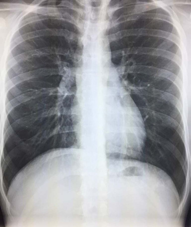
Bullae
The term “bullae” can be used in radiology reports of the chest. These air-filled spaces in the lungs, larger than one centimeter in diameter. Bullae appear as dark, thin-walled areas on lung imaging and can impact lung function. This article explains what bullae are, how they appear on different imaging tests, their causes, and what they might mean for your health.
What are Bullae?
Bullae are air-filled spaces within the lung tissue that measure more than 1 centimeter in diameter. These air pockets form when there is damage to the alveoli, the tiny air sacs responsible for oxygen exchange. When these air sacs break down and merge, they create larger air spaces called bullae. These structures can vary in size from just over a centimeter to sometimes occupying large portions of the lung.
Common Causes of Bullae
Bullae typically develop as a result of lung damage. The most common causes include:
Smoking is the leading cause of bullae formation. The toxic substances in cigarette smoke damage lung tissue over time, leading to the breakdown of alveolar walls and the formation of these air spaces. Long-term smokers are particularly susceptible to developing bullae.
Chronic obstructive pulmonary disease (COPD) is strongly associated with bullae. As COPD progresses, it causes emphysema, a condition where the alveoli are damaged and enlarged. These damaged air sacs can eventually form bullae.
Alpha-1 antitrypsin deficiency is a genetic condition that predisposes individuals to developing emphysema and bullae at a younger age, even without significant smoking history.
Inhalation of harmful substances such as coal dust, silica, or other industrial pollutants can also lead to bullae formation through chronic lung damage.
How Bullae Appear on Imaging
Bullae have distinctive appearances on various imaging modalities:
Chest X-rays
On a standard chest X-ray, bullae appear as thin-walled, air-filled spaces within the lung tissue. They are typically seen as dark areas with well-defined borders. However, smaller bullae may be difficult to detect on X-rays alone, particularly if they are surrounded by normal lung tissue or obscured by other structures.
CT Scanning
Computed tomography (CT) scans provide much more detailed imaging of bullae than X-rays. On CT scans, bullae appear as well-defined, air-filled spaces with thin walls. The higher resolution of CT allows for detection of smaller bullae and provides better assessment of their size, location, and relationship to surrounding lung tissue.
High-Resolution CT (HRCT)
HRCT is particularly valuable for evaluating bullae and other lung pathologies. This specialized form of CT provides detailed images of lung structure, allowing radiologists to:
- Precisely measure the size and number of bullae
- Assess the thickness of their walls
- Determine if there is any fluid or tissue within the bullae
- Evaluate surrounding lung tissue for additional abnormalities
Clinical Significance of Bullae
The presence of bullae on imaging studies has several important clinical implications:
Respiratory Function Impact
Bullae can significantly impact lung function. These air-filled spaces do not participate in gas exchange and essentially represent “dead space” within the lungs. Large or numerous bullae can reduce the available lung tissue for oxygen transfer, leading to decreased respiratory function and exercise tolerance.
Risk of Pneumothorax
One of the most serious complications associated with bullae is spontaneous pneumothorax, the sudden collapse of a lung. Bullae can rupture, allowing air to escape into the pleural space between the lung and chest wall. This complication often requires immediate treatment.
Infection Risk
Bullae can sometimes become infected, leading to a condition called an infected bulla or, if it forms an air-fluid level, a lung abscess. Infected bullae can be serious complications requiring antibiotic therapy and sometimes drainage or surgical intervention.
Treatment Considerations for Bullae
When bullae are identified on imaging, several treatment approaches may be considered:
Conservative Management
For many patients with small, asymptomatic bullae, regular monitoring may be the only intervention needed. This typically involves periodic imaging to assess for any growth or complications.
Smoking Cessation
For smokers with bullae, quitting smoking is essential to prevent further lung damage and bullae formation. Smoking cessation has been shown to slow the progression of bullous disease.
Surgical Options
For large bullae that cause significant symptoms or complications, surgical intervention may be necessary. Procedures include:
- Bullectomy: Surgical removal of bullae
- Lung volume reduction surgery: Removal of damaged lung tissue to improve function
- Lung transplantation: For severe cases with extensive bullous disease
Differential Diagnosis for Bullae
Several other lung conditions can sometimes be confused with bullae on imaging:
Pneumatoceles are air-filled spaces that typically form after infection or trauma and are usually temporary, unlike bullae which are permanent.
Blebs are small air-filled spaces (less than 1 cm) that form within the visceral pleura.
Cysts have a thicker wall than bullae and may contain fluid or solid material rather than just air.
Conclusion
Bullae can impact respiratory function and increase the risk of complications such as pneumothorax. Imaging tests provide important diagnostic information about bullae and underlying lung conditions such as emphysema and COPD. Understanding the appearance, causes, and clinical significance of bullae is important for patients and their doctors for optimal patient care.
References
- https://my.clevelandclinic.org/health/diseases/24728-bullous-emphysema
- https://www.ncbi.nlm.nih.gov/books/NBK537243/
- https://learningradiology.com/notes/chestnotes/bullous%20dz.htm

