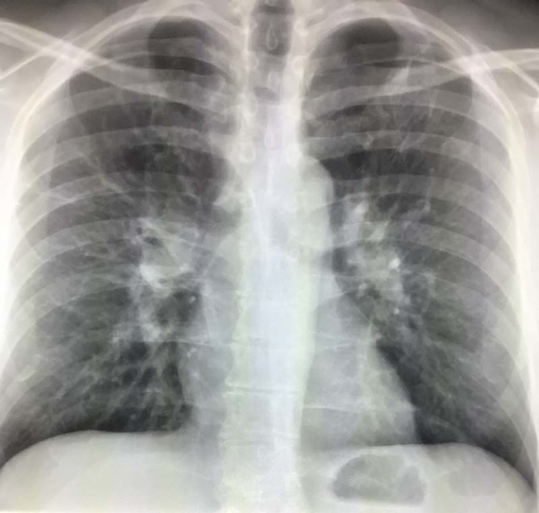
CT of the Chest for Chest Pain
CT is often done to diagnose the cause of chest pain. CT is good at diagnosing some of the benign and life threatening conditions that can cause chest pain. Chest pain can be caused by many different conditions arising from the heart, blood vessels of the chest, esophagus, chest wall and upper abdomen.
What are some of the conditions we can diagnose on CT?
Diseases of the lungs can cause chest pain. Pneumonia or infection can be diagnosed with CT as an area of white amidst the dark of the normal lungs. A collapsed lung or pneumothorax can be seen as air around the lung and collapse of the lung lobe. Fluid around the lung is called a pleural effusion and can be well seen. Cancer involvement of the chest can also be diagnosed.
Diseases of the blood vessels of the chest can cause chest pain. Aortic dissection which is a tear of the inner lining of the aorta splits the aorta into two lumens. Aortic aneurysms can be readily diagnosed. Clot to the lungs can be diagnosed with a specialized CT called an angiogram. Clots most commonly arise from the leg veins.
Heart disease is not well seen on routine chest CTs.
Diseases of the heart are not well diagnosed with CT. We can see fluid around the heart called pericardial effusion. We can’t see diseases of the heart valves on routine CTs. We can’t diagnose heart attack or blocked coronaries on routine CTs. Some specialized CTs can image the coronary arteries which when blocked lead to heart attack. Angiograms and Echocardiograms or ultrasound of the heart is the best test to image the heart.
CT is not a great test for the esophagus
Diseases of the esophagus are not well diagnosed with CT. We can sometimes see foreign bodies or tears of the esophagus. We can also see masses arising from the esophagus when large. We usually can’t see inflammation of the esophagus, heart burn, or spasms. X-ray tests with swallowed barium and endoscopy are the best tests for the esophagus.
Chest wall conditions like a broken rib can be seen on CT. Costochondritis which is an inflamed cartilage of the rib where it meets the breast bone can not be seen on CT. We can see cancer when it spreads to the chest wall.
Upper abdominal inflammatory conditions of the gallbladder and pancreas to name a few can be seen on CT. Often we need a dedicated imaging test of the abdomen to diagnose these conditions.
Some psychiatric conditions like panic disorder and anxiety can cause chest pain. Often these patients will still get a big workup to exclude serious conditions. A good history can often make the diagnosis.
CT is an excellent test to exclude some important treatable and life threatening conditions that cause chest pain. But it is important to remember that many conditions that cause chest pain can not be seen on CT. Dangerous conditions of the heart can not usually be diagnosed with routine CT. A good clinical history, physical exam, blood tests and other imaging may be needed to arrive at the correct diagnosis.

