Interstitial Meaning
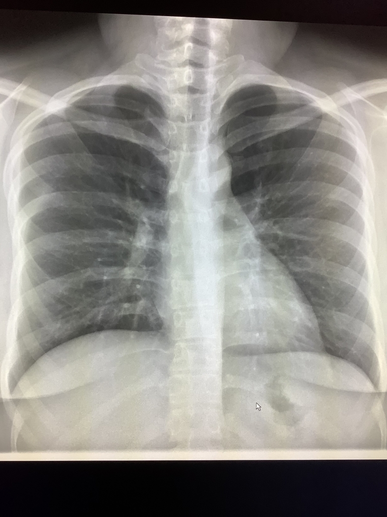
The term interstitial can appear in chest X-ray and CT scans reports. Interstitial abnormalities occur between the air spaces of the lung where carbon dioxide and oxygen exchange occurs. This article explains what interstitial findings mean in radiology and what kinds of abnormalities are commonly seen with this pattern.
What Does Interstitial Mean in Medical Imaging?
In medical terminology, “interstitial” refers to the spaces between structures in the body. Most commonly, when radiologists mention interstitial findings, they’re referring to changes in the tissue that exists between the air sacs (alveoli) in your lungs. These spaces contain a delicate network of blood vessels, connective tissue, and other supporting structures that are crucial for proper lung function.
Interstitial abnormalities appear as specific patterns on imaging that trained radiologists can identify. These patterns may show as lines, nodules, or areas of increased density that differ from the normal appearance of healthy lung tissue.
Common Interstitial Patterns on Imaging
When looking at imaging studies, radiologists identify several distinct interstitial patterns:
Reticular Pattern
A reticular pattern appears as a network of fine lines on imaging. This pattern resembles a mesh or net and typically indicates thickening of the interstitial tissue. On X-rays and CT scans, these show up as thin, intersecting lines throughout the lung fields.
Ground-Glass Opacity
Ground-glass opacity (GGO) is a hazy appearance on imaging that doesn’t obscure underlying lung structures. This pattern gets its name because it resembles ground glass – translucent but not completely transparent. Ground-glass opacities often indicate partial filling of air spaces or thickening of the interstitial tissue.
Nodular Pattern
A nodular interstitial pattern shows as multiple small, round opacities scattered throughout the lungs. Depending on their distribution, size, and other characteristics, nodular patterns can indicate various conditions from infection to inflammatory diseases.
Honeycombing
Honeycombing represents advanced interstitial lung disease. It appears as clustered cystic air spaces with well-defined walls, resembling a honeycomb. This finding often indicates irreversible lung damage and fibrosis.
What Causes Interstitial Changes?
Interstitial abnormalities can result from numerous conditions affecting the lungs. Some common causes include:
Interstitial Lung Diseases
Interstitial lung diseases (ILDs) comprise a large group of disorders characterized by inflammation and scarring of lung tissue. These include conditions like idiopathic pulmonary fibrosis, sarcoidosis, and hypersensitivity pneumonitis.
Infections
Certain infections, including viral pneumonia, can cause temporary interstitial changes. COVID-19, for example, commonly shows as ground-glass opacities on chest imaging.
Environmental Exposures
Exposure to certain substances like asbestos, silica dust, coal dust, or radiation can damage the interstitial spaces in the lungs, leading to visible changes on imaging.
Medication Effects
Some medications can cause drug-induced interstitial lung disease as a side effect. Chemotherapy drugs, certain antibiotics, and anti-inflammatory medications are known potential causes.
Heart Problems
Heart failure can lead to fluid buildup in the lungs, which may appear as interstitial changes on imaging. This pattern often shows more prominently in the lower portions of the lungs.
How Radiologists Evaluate Interstitial Findings
When radiologists spot interstitial changes on imaging, they consider several factors:
Distribution Pattern
The location of abnormalities provides important diagnostic clues. Some conditions typically affect the upper lungs, while others predominantly involve lower or peripheral regions.
Symmetry
Symmetrical (affecting both lungs equally) versus asymmetrical changes can help narrow down potential causes.
Associated Findings
Other findings that accompany interstitial changes, such as enlarged lymph nodes, pleural effusions (fluid around the lungs), or changes in heart size, provide additional diagnostic information.
Progression Over Time
Comparing current imaging with previous studies helps assess whether interstitial changes are stable, improving, or worsening.
What Happens After Interstitial Findings Are Detected?
If your imaging shows interstitial abnormalities, your doctor might recommend:
Additional Imaging
High-resolution CT scans provide more detailed images of interstitial changes than standard X-rays and can help better characterize the abnormalities.
Pulmonary Function Tests
These breathing tests measure how well your lungs work and can detect abnormalities even before symptoms appear.
Bronchoscopy
This procedure allows doctors to look inside your airways and collect tissue samples if needed.
Surgical Lung Biopsy
In some cases, a small piece of lung tissue may need to be removed and examined under a microscope to determine the exact cause of interstitial changes.
When to Be Concerned About Interstitial Findings
Not all interstitial findings indicate serious disease. Some may represent normal aging changes or remnants of previous infections that have healed. However, certain characteristics raise concern:
- Progressive worsening of findings over time
- Extensive involvement throughout both lungs
- Association with significant symptoms like shortness of breath
- Presence of honeycombing, which suggests irreversible fibrosis
In my practice as a radiologist, interstitial changes often indicate an active process requiring follow-up when they appear as new findings or show progression compared to prior studies.
Conclusion
Interstitial findings on radiology reports refer to abnormalities in the supporting tissue between air sacs in the lungs. These findings appear as distinctive patterns on imaging studies that radiologists can identify and characterize. While some interstitial changes may represent benign or temporary conditions, others indicate chronic or progressive lung diseases requiring treatment. Understanding the meaning of interstitial abnormalities can help you have more informed discussions with your doctor about your imaging results and any recommended follow-up.
References

