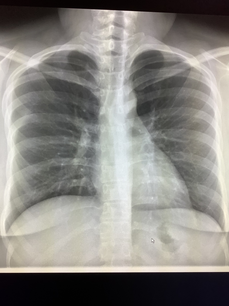
Pneumomediastinum Symptoms, Diagnosis, Imaging and Treatment
Pneumomediastinum is a condition that may sound complex, but it involves air getting trapped in the mediastinum, the space in your chest between the lungs. It can cause discomfort and a range of symptoms. In this article, we’ll dive into the symptoms, diagnosis, and treatment options for pneumomediastinum, with a particular focus on imaging techniques.
Symptoms of Pneumomediastinum
Recognizing the symptoms of pneumomediastinum is crucial for early diagnosis and treatment. Common symptoms include:
- Chest Pain: One of the primary indicators is chest pain, often described as sharp or stabbing. It can radiate to the neck or back.
- Shortness of Breath: Individuals with pneumomediastinum may experience difficulty breathing or a sensation of not getting enough air.
- Subcutaneous Emphysema: This is a unique symptom where air escapes into the tissues under the skin, causing a crackling or popping sensation when touched.
- Difficulty Swallowing: Some people may find it hard to swallow food or liquids.
- Voice Changes: Pneumomediastinum can affect the vocal cords, leading to hoarseness or a change in voice.
Diagnosis of Pneumomediastinum
When these symptoms arise, it’s crucial to seek medical attention promptly. Your healthcare provider will perform various tests to confirm pneumomediastinum. Some diagnostic tools include:
- Chest X-Ray: This is often the first imaging technique used to visualize the air in the mediastinum. It’s a quick and effective way to identify pneumomediastinum.
- Computed Tomography (CT) Scan: A CT scan provides more detailed images, helping to determine the extent and cause of the air accumulation.
- Esophagography: Sometimes, a barium swallow test may be performed to rule out other conditions and confirm pneumomediastinum.
Imaging in Pneumomediastinum
Imaging plays a pivotal role in diagnosing and understanding pneumomediastinum. Among the various techniques, CT scans are particularly valuable. These scans allow healthcare professionals to obtain cross-sectional images of the chest, providing detailed information about the location and extent of the air accumulation.
CT scans can also help identify potential underlying causes, such as a ruptured air sac or damage to the esophagus or trachea. This information is crucial for determining the most appropriate treatment plan.
Treatment Options for Pneumomediastinum
The treatment of pneumomediastinum depends on its underlying cause and severity. Here are some common approaches:
- Observation: In mild cases, pneumomediastinum may resolve on its own with conservative management. Regular monitoring and follow-up appointments are essential.
- Oxygen Therapy: Oxygen therapy may be used to relieve symptoms and facilitate the absorption of the trapped air.
- Surgery: In severe cases or when there’s a significant underlying issue, surgical intervention may be necessary to repair the damage.
- Treatment of Underlying Cause: Treating the root cause of pneumomediastinum, such as addressing a ruptured air sac or esophageal injury, is critical for a successful outcome.
Conclusion
Pneumomediastinum can cause distressing symptoms, but early diagnosis and appropriate treatment can lead to a full recovery. Imaging techniques, particularly CT scans, play a crucial role in identifying the condition’s extent and underlying causes.
If you experience symptoms like chest pain, shortness of breath, or subcutaneous emphysema, don’t hesitate to seek medical attention. Your healthcare provider can use these imaging tools to diagnose pneumomediastinum accurately and develop an effective treatment plan tailored to your specific needs.

