Pneumothorax On Chest X-ray
A pneumothorax or collapsed lung is a diagnosis which can be made on a chest X-ray. This potentially life threatening condition needs a prompt diagnosis and treatment.
What is a pneumothorax?
A pneumothorax is air leaking from the lung into the space between the lung and chest wall. This will cause the lung to collapse to varying degrees.
What causes a pneumothorax?
A pneumothorax can happen spontaneously in a person with healthy lungs or in someone who has lung disease.
Patients who have lung disease such as emphysema and cystic fibrosis can predispose someone to develop a pneumothorax.
Lung cancer, abscess and pneumonia are some other conditions which can predispose a patient to a pneumothorax.
Patients who have had a medical procedure such as a lung biopsy can develop a pneumothorax.
Both blunt and penetrating trauma can result in pneumothorax. It is important to make sure that there is no pneumothorax when a patient breaks a rib.
Symptoms of pneumothorax
A pneumothorax can be asymptomatic or associated with severe breathlessness, chest pain and low blood pressure.
Is a pneumothorax dangerous? (or cancerous)
If the pneumothorax is associated with increasing pressure on the adjacent chest structures like the heart, then this can be life threatening and needs prompt treatment.
A pneumothorax can cause breathlessness, chest pain and rapid heart rate. A pneumothorax can enlarge in some cases.
A pneumothorax can rarely be a presentation of cancer. CT imaging and biopsies may be needed for the diagnosis.
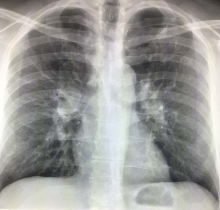
What does pneumothorax look like on a chest x-ray?
A pneumothorax on chest X-ray is diagnosed by seeing the upper edge of the collapsed lung and absence of normal lung above it. The space between the collapsed lung and chest wall is filled with air. An estimate of the size of the pneumothorax is made by the radiologist to assist with treatment.
Are there other tests for pneumothorax?
In some cases, additional X-ray views may be necessary to help detect the pneumothorax. These may include X-rays where you take a breathe in and out or lay on your side. Sometimes a pneumothorax can only be seen on a CT scan.
Can other conditions look like pneumothorax on chest X-ray?
There are mimics of pneumothorax on chest X-ray. Skin folds can sometimes project onto the lung and mimic a pneumothorax.
The edge of a bone such as the scapula project onto the lung and give a similar appearance.
Lack of lung markings can be seen with emphysema. This can sometimes give the appearance of a pneumothorax.
Further imaging with CT will help with confusing cases.
Pneumothorax treatment
The decision to observe or treat a pneumothorax depends on the size, symptoms and any underlying lung disease.
Your physician may choose to observe the pneumothorax with serial X-rays, place a needle into the chest to remove the air or insert a drain.
In certain cases, a pleuordesis may be performed where the lung is fixed to the chest wall. This will prevent future recurrence of pneumothorax.
Pneumothorax on chest x-ray: summary
Pneumothorax is a potentially life threatening condition which is often diagnosed on X-ray. Some pneumothoraces are observed and others treated. CT imaging can help search for an underlying cause.