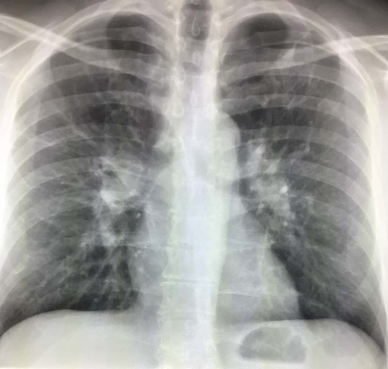
Chest Ultrasound
Chest ultrasounds have emerged as an important tool in medical imaging, offering a non-invasive method to visualize the chest. This technique is particularly useful for diagnosing and monitoring various conditions affecting the heart, lungs, and other areas within the thorax. By employing sound waves to create images, chest ultrasounds provide a safe alternative to other imaging modalities.
Understanding Chest Ultrasound
A chest ultrasound, also known as a thoracic ultrasound, is a diagnostic procedure that uses high-frequency sound waves to produce images of the chest’s internal structures. Unlike X-rays and CT scans, which use ionizing radiation, ultrasounds are radiation-free and are considered safer, especially for pregnant women and young children. The procedure is typically quick, painless, and can be performed at the bedside, making it highly accessible for critically ill or immobile patients.
Benefits of Chest Ultrasound
The advantages of chest ultrasounds are manifold. They provide detailed images of the chest cavity, including the heart, lungs, and the area surrounding these organs. This imaging technique is especially useful for detecting fluid around the heart (pericardial effusion), fluid in the lungs (pleural effusion), and abnormalities in the lung tissue itself. Furthermore, chest ultrasounds can guide procedures such as needle biopsies or fluid drainage, enhancing their safety and accuracy.
How Chest Ultrasound Works
During a chest ultrasound, a small, hand-held device called a transducer is used. This device sends out high-frequency sound waves that bounce off the body’s tissues and organs. The echoes are then captured and transformed into real-time images displayed on a monitor. The procedure usually requires the patient to be positioned in a way that allows the best possible access to the chest area being examined. The process is quick, typically taking around 30 minutes to complete, and does not require any special preparation.
Applications of Chest Ultrasound
Chest ultrasounds have a wide range of applications. They are particularly effective in evaluating symptoms such as chest pain, shortness of breath, and persistent cough. By providing real-time images, they allow healthcare providers to assess the movement and function of the heart and lungs. Additionally, chest ultrasounds play a critical role in emergency medicine, where they are used to quickly diagnose conditions like cardiac tamponade (compression of the heart due to fluid accumulation).
Comparing Chest Ultrasound with Other Imaging Techniques
When compared to other imaging techniques such as X-rays and CT scans, chest ultrasounds offer the distinct advantage of being radiation-free. They are also more accessible and cost-effective, making them an excellent option for initial evaluations or in settings where other imaging modalities are not available. However, it’s important to note that while chest ultrasounds are highly effective for examining soft tissues and fluid, they may not be as effective for visualizing bones or certain lung conditions where other imaging techniques might be preferred.
Preparing for a Chest Ultrasound
Preparing for a chest ultrasound is straightforward. There are generally no special preparations required, but patients may be advised to wear comfortable, loose-fitting clothing. They might need to remove clothing and jewelry from the waist up and wear a gown during the procedure. Following the ultrasound, patients can usually return to their normal activities immediately, as there are no side effects or recovery time associated with the procedure.
Conclusion
Chest ultrasounds serve as an important tool in modern medicine, offering a safe, efficient, and non-invasive way to visualize the chest’s internal structures. With benefits ranging from real-time imaging to the absence of radiation exposure, it’s no wonder that this imaging technique has become important in diagnosing and monitoring a wide array of chest conditions. Whether it’s guiding emergency procedures or evaluating chronic symptoms, the versatility and accessibility of chest ultrasounds make them an important part of patient care.

