Kerley B Lines
Kerley B lines are specific radiographic findings visible on chest X-rays that represent thickened interlobular septa in the lungs. These short horizontal lines appear primarily at the lung periphery and indicate fluid or cellular infiltration in the lung’s connective tissue. Commonly associated with conditions such as pulmonary edema, interstitial lung disease, and lymphangitic carcinomatosis. This article explains what Kerley B lines represent, their clinical significance, and what patients should understand when these findings appear on their radiology reports.
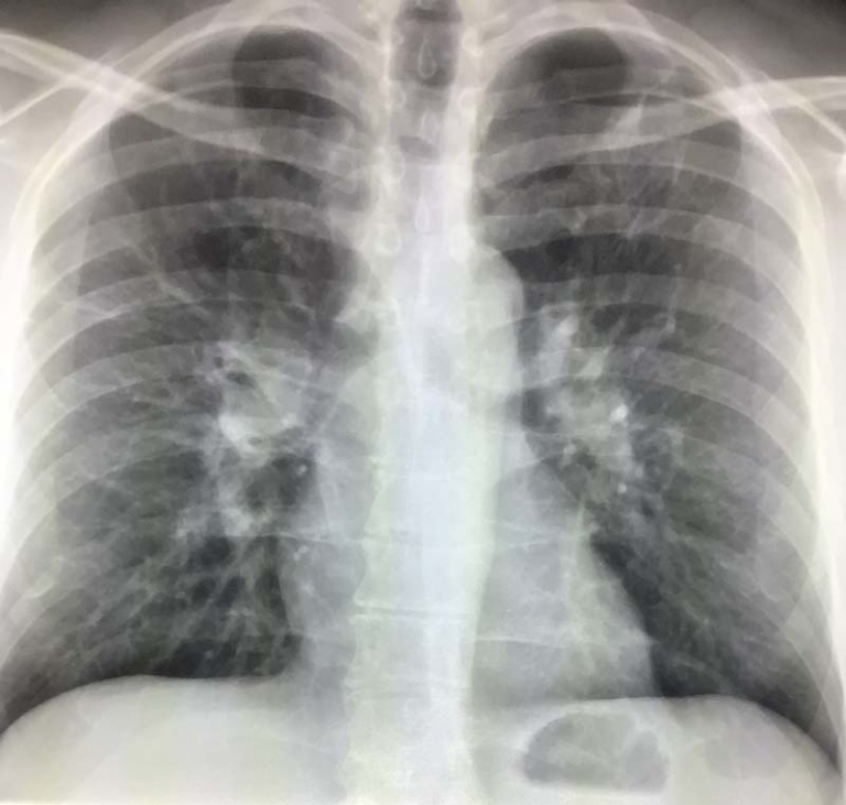
What Are Kerley B Lines?
Kerley B lines appear as short, horizontal, linear opacities primarily visible at the edges of the lungs on chest X-rays. These lines are typically 1-2 cm in length and are most commonly seen at the lung bases, extending to the pleural surface (the thin membrane covering the lungs). These lines represent thickened interlobular septa – the boundaries between small sections of lung tissue.
While these lines may appear insignificant to untrained eyes, for radiologists and pulmonologists, they provide valuable diagnostic information about what might be happening inside your lungs.
Common Causes of Kerley B Lines
Kerley B lines aren’t a disease themselves but rather a radiographic sign of underlying conditions. The most frequent causes include:
Pulmonary Edema
Pulmonary edema – fluid accumulation in the lung tissues – is the most common reason Kerley B lines appear on X-rays. This condition often results from heart failure, where the heart cannot effectively pump blood, leading to fluid backing up into the lungs. The lines become visible as the interlobular septa fill with fluid.
Interstitial Lung Disease
Various interstitial lung diseases can cause Kerley B lines to appear. These diseases involve inflammation and scarring of the lung tissue, which thickens the interlobular septa and creates visible lines on X-rays.
Lymphangitic Carcinomatosis
This serious condition involves cancer spreading through the lymphatic vessels of the lungs. As tumor cells infiltrate the lymphatic vessels that run within the interlobular septa, they cause thickening that appears as Kerley B lines.
How Kerley B Lines Are Detected
Radiologists identify Kerley B lines during the examination of chest X-rays. While these lines can sometimes be challenging to spot, they become more prominent in certain conditions:
Chest X-ray Findings
On standard chest X-rays, Kerley B lines appear as thin horizontal lines at the lung periphery, usually measuring between 1-2 cm in length. They’re most visible at the costophrenic angles (where the diaphragm meets the chest wall) and lateral lung bases.
Advanced Imaging Techniques
While conventional X-rays remain the primary method for detecting Kerley B lines, high-resolution CT scans can provide more detailed images of interlobular septal thickening. CT scans may detect these changes even when they’re not yet visible on standard X-rays.
Clinical Significance of Kerley B Lines
The presence of Kerley B lines often indicates a significant underlying condition that requires medical attention:
Indicator of Heart Failure
In patients with heart problems, Kerley B lines often represent pulmonary edema due to left-sided heart failure. Their appearance might precede other symptoms, making them a valuable early warning sign.
Monitoring Disease Progression
For patients with known interstitial lung disease or heart failure, the appearance or disappearance of Kerley B lines helps clinicians assess treatment effectiveness and disease progression.
Differential Diagnosis
The pattern and distribution of Kerley B lines, along with other radiographic findings, help physicians narrow down possible diagnoses. For instance, lines predominantly in the lower lung zones often suggest heart failure, while a more diffuse pattern might indicate interstitial lung disease.
Treatment Approaches
Treatment for conditions causing Kerley B lines focuses on addressing the underlying cause:
Managing Heart Failure
For heart-related causes, treatment typically includes medications to reduce fluid retention and improve heart function, such as diuretics, ACE inhibitors, and beta-blockers.
Treating Interstitial Lung Disease
Treatment may involve anti-inflammatory medications, immunosuppressants, or antifibrotic drugs depending on the specific type of interstitial lung disease.
Cancer Treatment
For lymphangitic carcinomatosis, treatment targets the underlying cancer, potentially including chemotherapy, radiation, or immunotherapy.
When to Seek Medical Attention
If your radiology report mentions Kerley B lines, it’s important to discuss this finding with your healthcare provider. While not always indicating an emergency, these lines suggest an underlying condition that requires evaluation and potentially treatment.
Symptoms that might accompany conditions associated with Kerley B lines include:
- Shortness of breath, especially when lying flat
- Persistent dry cough
- Unexplained fatigue
- Swelling in the legs or ankles
- Rapid weight gain due to fluid retention
Long-term Outlook
The prognosis for patients with Kerley B lines varies widely depending on the underlying cause. Many heart failure patients experience resolution of these lines with appropriate treatment, while those with advanced interstitial lung disease or metastatic cancer may have a more guarded outlook.
Conclusion
Kerley B lines represent a radiographic finding that provides diagnostic information about potential heart and lung conditions. By recognizing the significance of these subtle lines, radiologists can help identify conditions at earlier, more treatable stages. Your doctor will best know how to manage the radiologic findings in your specific case.
References
- https://radiopaedia.org/articles/septal-lines-in-lung-1?lang=us
- https://introductiontoradiology.net/courses/rad/cxr/pathology2Cchest.html
- https://thecommonvein.com/heart/kerley-lines/